
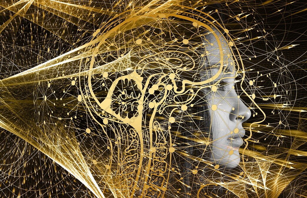
This is a multi-disciplinary approach that is inspired by biological evolution, quantum biology, and the human skin cell from the Eye of Unity Foundation. Evolutionary biology suggests that animals that appear similar in appearance have similar dna sequences. Quantum biology suggests that the way in which molecules react to light is a function of their dna sequence. Furthermore, the human skin cell is a model of the whole human body and can be used to study the function of various genes.
A key challenge in this area is the fact that fluorescence is an unusual and spontaneous property acquired by only a few types of organisms. The evolution of fluorescence in animals is a fascinating and important story, and CRSPR aims to study this process. One of the most exciting developments in understanding the evolution of fluorescence is the discovery that many different fluorescent proteins can be expressed in the genome of animals. These proteins can be inserted into cells and expressed in many different ways, allowing researchers to study and manipulate the mechanisms of many different kinds of cells in the body to see how fluorescence is used in different biological processes.
What is CRSPR?
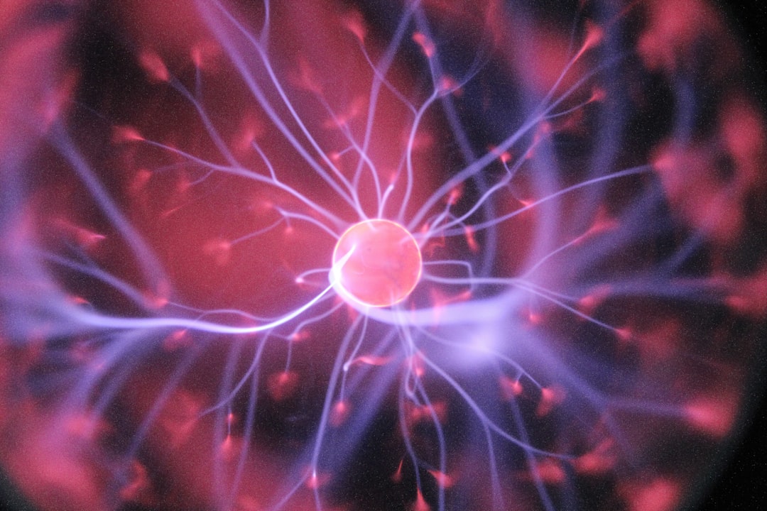
CRISPR is a gene editing technology that has been making waves in the scientific community for its ability to edit DNA with unparalleled precision used in conjunction with Quantum Biology. One of the first applications of quantum biology was the development of green fluorescent protein, or GFP. GFP is a protein that glows green under certain conditions. It was first discovered in jellyfish, and has since been used in a variety of applications, including genetic engineering and molecular biology. Welcome to the future. Thanks to quantum biology, we now have the ability to edit skin cell DNA with unprecedented accuracy. This could lead to all sorts of amazing applications, such as fluorescent skin cells and regenerative medicine.
CRSPR is a year-long program that aims to honor the life of Prof. H.L. Cross, who is known as the “father of fluorescence in biology”. His research focused on the use of organic fluorophores (including fluorescein) to study the dynamics of living biological systems. Fluorescent proteins are proteins that glow when light is shone on them, but they can also be genetically modified to emit different colors of light. This type of fluorescence is an example of organic fluorescence, which is the evolution of fluorescence in animals.
Editing the Skin-Cell DNA with CRSPR.
To edit the skin-cell dna, you need to use a fluorescent protein-based reporter molecule that can be inserted into the DNA through the CRISPR/Cas9 system. The first step is to create a cut in the DNA with CRISPR/Cas9; next, the fluorescent protein needs to be inserted into the DNA. Once these two steps have been completed, the fluorescent protein expression needs to be turned on in the skin cell. This is done by using a “switch” molecule, which is a molecule that is able to turn the fluorescent protein on and off based on the cellular environment.
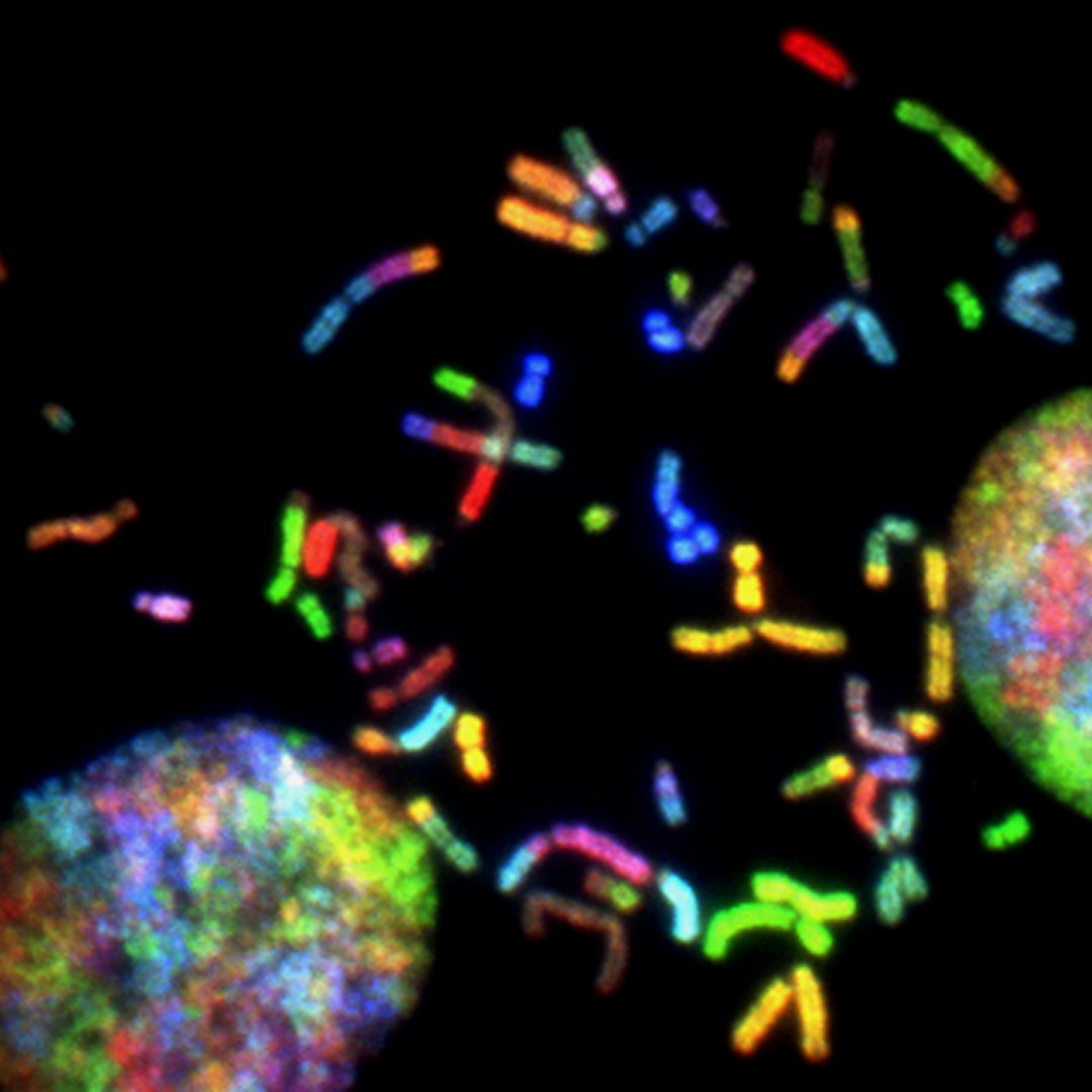
The cell’s DNA is a double-helix of two strands—one of which is the DNA-molecule, and the other of which is the RNA-molecule. At the center of each strand, there are proteins known as histones, which act as a physical basis for the DNA-molecule. In most cells, the histones are in a tight, coiled-up ball, known as a nucleosome. However, in the single-cell, the histones are much looser, and this allows the DNA-molecule to be more easily accessible to the DNA-replication machinery.
A laser is used to alter the DNA of skin cells, which are then fixed on a slide and viewed under the microscope. The laser will create a hole in the cell’s membrane, which allows the DNA to be exposed. This hole is then filled with a fluorescent dye which will ‘tell’ you where the DNA is located. The laser is also used to ‘cut’ the DNA out and send it to you. After that, you can use a computer program to find the spot on the DNA that you want to change.
Building Blocks and Components of The Single Cell
This toolkit is a very useful tool for understanding how the single-cell works, and it’s made up of an assortment of machines.
- Cell Membrane
The first of which is the cell’s membrane. The cell’s membrane is like a big rubber-band that holds the cell inside the body. It plays a key role in the cell’s division, as well as its metabolism.
- THE CHEMICAL COMPOUND, ATP.
ATP is a chemical compound that’s commonly used by cells as a source of energy. In its free form, it’s like a big chemical battery that the cell can use as a source of energy.
- THE PROTEIN, HISTONE.
HISTONE is a protein that’s found in the nucleus of the cell.
It’s like a big ball of coiled-up protein, and it acts as a physical basis for the DNA-molecule.
- The RNA-molder.
RNA is a compound that contains a series of chemical chains that are strung together in a complex fashion. The RNA-molecule is used as a messenger of sorts, and it helps the cell to translate the DNA-molecule’s instructions into proteins.
- The replication machine.
A replication machine is a chemical machine that is used by the cell to copy the DNA-molecule.
It’s used to make a new copy of the DNA, and it’s made up of many different parts, including:
- DNA polymerase.
DNA polymerase is a type of enzyme that helps the cell to copy the DNA-molecule.
- The ribosome.
The ribosome is a protein machine that is used by the cell to translate the RNA-molecule’s instructions into proteins.
- The chromatin remodeling machine.
The chromatin remodeling machine is a protein machine that is used by the cell to change the structure of the DNA.
It’s used to change the DNA’s structure from a coiled-up ball to a more open, accessible structure.

Evolution and The Three Parts of The Fluorescent Single Cell
The evolution of fluorescence in animals can be traced back to 600 million years ago when animals began to use fluorescence to make their skin and muscles easier to see in the dark. Subsequently, these fluorescence-emitting proteins evolved into a wide range of functions, and today they are found in all types of life.
Some organisms also use fluorescence to make their sex organs easier to recognize in the dark. In this case, the proteins that fluoresce are known as ‘fluorescent proteins’ and they are made by a particular group of bacteria called the cyanobacteria. These bacteria use fluorescence to transmit signals to other bacteria and even to other organisms. The human skin cell has its own fluorescent proteins, and scientists are now using these to study the structure of skin cells.
- The nucleus.
The nucleus is the biggest organelle in the cell, and it’s also considered a cell’s “brain,” as it contains all of the cell’s DNA.The cytoplasm.
- The cytoplasm
The cytoplasm is a liquid-like substance that’s filled with many different types of proteins, and it’s used to perform many different functions.
- The mitochondria.
The mitochondria are organelles that are found in the cell’s cytoplasm, and they help the cell to produce energy.
The mitochondria are important, because they play a role in the cell’s metabolism.